
Researchers show the importance of the liver-brain axis in Alzheimer'sdisease UAB_info
]. This is consistent with our histopathological and biochemical results for NTg animals and becomes evident with the correlations between the liver index, enzymatic activity of antioxidant enzymes, and levels of pro-inflammatory factors. Despite not presenting with this liver disease, 3xTg-AD animals were found to be affected by a group of diseases known as amyloidosis. Based on these results, oxidative stress and inflammation were found to be exacerbated.
Hepatic dysfunction was determined at the organ, tissue, and cellular levels in 16-month-old animals. The hallmarks of 3xTg-AD liver dysfunction included hepatomegaly, acute amyloidosis, and ballooning. In addition, increased oxidative stress and inflammation were shown as the dysregulation of antioxidant enzymes and increased levels of pro-inflammatory cytokines, with a sexual dimorphism.
Neophobia has previously been shown to exert a clear psychological effect by increasing hyperactivity, anxiogenic patterns, and bizarre/flight behavior. In this study, neophobia correlated with oxidative stress variables in the liver.
Australia Latest News, Australia Headlines
Similar News:You can also read news stories similar to this one that we have collected from other news sources.
 Progressive inflammation reduces high-frequency EEG activity and cortical dendritic arborisation in late gestation fetal sheep - Journal of NeuroinflammationBackground Antenatal infection/inflammation is associated with disturbances in neuronal connectivity, impaired cortical growth and poor neurodevelopmental outcomes. The pathophysiological substrate that underpins these changes is poorly understood. We tested the hypothesis that progressive inflammation in late gestation fetal sheep would alter cortical neuronal microstructure and neural function assessed using electroencephalogram band power analysis. Methods Fetal sheep (0.85 of gestation) were surgically instrumented for continuous electroencephalogram (EEG) recording and randomly assigned to repeated saline (control; n = 9) or LPS (0 h = 300 ng, 24 h = 600 ng, 48 h = 1200 ng; n = 8) infusions to induce inflammation. Sheep were euthanised 4 days after the first LPS infusion for assessment of inflammatory gene expression, histopathology and neuronal dendritic morphology in the somatosensory cortex. Results LPS infusions increased delta power between 8 and 50 h, with reduced beta power from 18 to 96 h (P | 0.05 vs. control). Basal dendritic length, numbers of dendritic terminals, dendritic arborisation and numbers of dendritic spines were reduced in LPS-exposed fetuses (P | 0.05 vs. control) within the somatosensory cortex. Numbers of microglia and interleukin (IL)-1β immunoreactivity were increased in LPS-exposed fetuses compared with controls (P | 0.05). There were no differences in total numbers of cortical NeuN + neurons or cortical area between the groups. Conclusions Exposure to antenatal infection/inflammation was associated with impaired dendritic arborisation, spine number and loss of high-frequency EEG activity, despite normal numbers of neurons, that may contribute to disturbed cortical development and connectivity.
Read more »
 Associations between circulating cell-free mitochondrial DNA, inflammatory markers, and cognitive and physical outcomes in community dwelling older adults - Immunity & AgeingBackground Dementia and frailty are common age-related syndromes often linked to chronic inflammation. Identifying the biological factors and pathways that contribute to chronic inflammation is crucial for developing new therapeutic targets. Circulating cell-free mitochondrial DNA (ccf-mtDNA) has been proposed as an immune stimulator and potential predictor of mortality in acute illnesses. Dementia and frailty are both associated with mitochondrial dysfunction, impaired cellular energetics, and cell death. The size and abundance of ccf-mtDNA fragments may indicate the mechanism of cell death: long fragments typically result from necrosis, while short fragments arise from apoptosis. We hypothesize that increased levels of necrosis-associated long ccf-mtDNA fragments and inflammatory markers in serum are linked to declines in cognitive and physical function, as well as increased mortality risk. Results Our study of 672 community-dwelling older adults revealed that inflammatory markers (C-Reactive Protein, soluble tumor necrosis factor alpha, tumor necrosis factor alpha receptor 1 [sTNFR1], and interleukin-6 [IL-6]) positively correlated with ccf-mtDNA levels in serum. Although cross-sectional analysis revealed no significant associations between short and long ccf-mtDNA fragments, longitudinal analysis demonstrated a connection between higher long ccf-mtDNA fragments (necrosis-associated) and worsening composite gait scores over time. Additionally, increased mortality risk was observed only in individuals with elevated sTNFR1 levels. Conclusion In a community dwelling cohort of older adults, there are cross-sectional and longitudinal associations between ccf-mtDNA and sTNFR1 with impaired physical and cognitive function and increased hazard of death. This work suggests a role for long ccf-mtDNA as a blood-based marker predictive of future physical decline.
Read more »
 Warning as ‘silent’ but deadly disease on the rise - the 3 signs you must knowCASES of a deadly non-alcoholic fatty liver disease have been soaring over the past three decades, according to a new study. The condition, which causes inflammation of the organ, can lead to liver…
Read more »
 Newly discovered genetic defect disrupts blood formation and immune systemIn the quest to find the origin of the puzzling symptoms in four children, researchers from St. Anna Children's Cancer Research Institute, the CeMM Research Center for Molecular Medicine of the Austrian Academy of Sciences (ÖAW), and the Medical University of Vienna have discovered a completely new disease, linking disruptions of blood formation, the immune system, and inflammation. This discovery provides the basis for a better understanding of similar diseases. It is a milestone that the researchers have now published in the New England Journal of Medicine.
Read more »
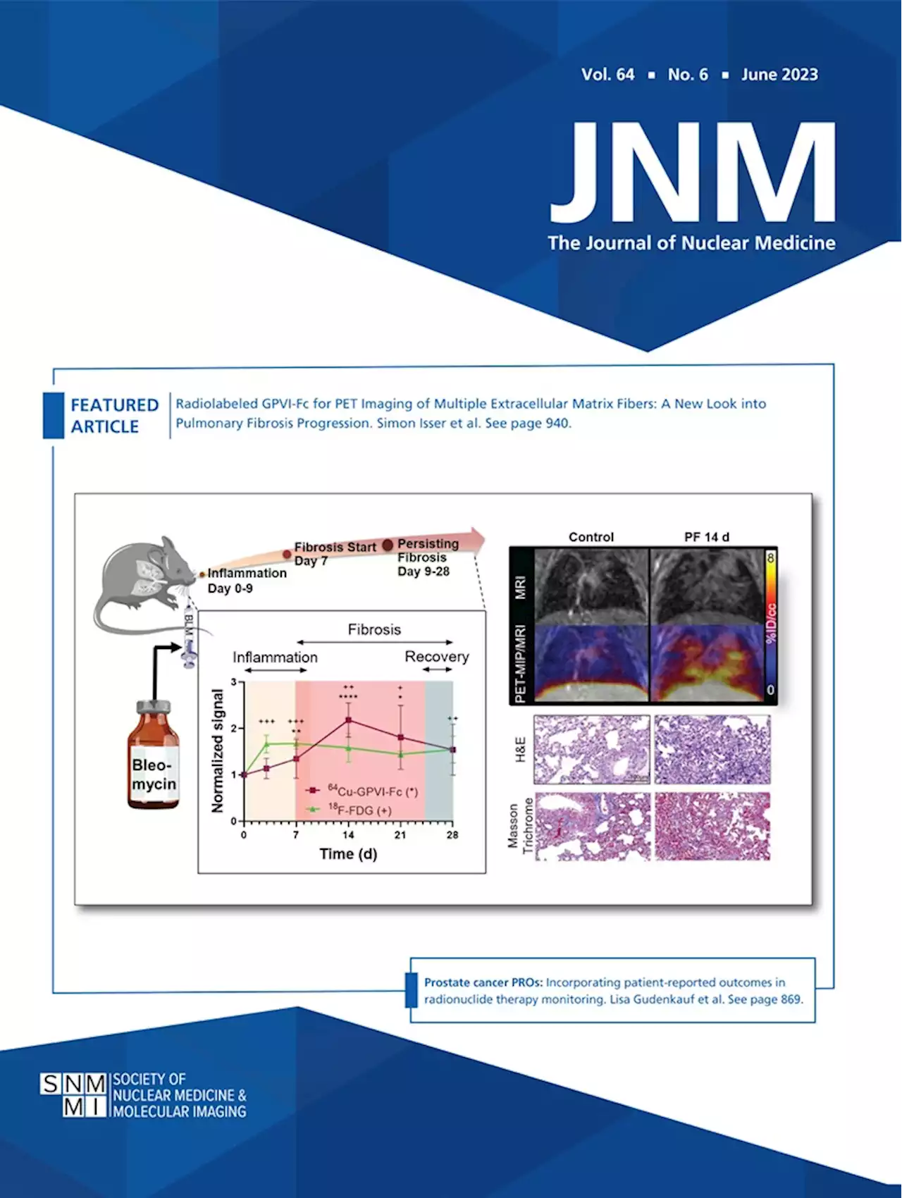 Radiolabeled GPVI-Fc for PET Imaging of Multiple Extracellular Matrix Fibers: A New Look into Pulmonary Fibrosis ProgressionInvariably fatal and with a particularly fast progression, pulmonary fibrosis (PF) is currently devoid of curative treatment options. Routine clinical diagnosis relies on breathing tests and visualizing the changes in lung structure by CT, but anatomic information is often not sufficient to identify early signs of progressive PF. For more efficient diagnosis, additional imaging techniques were investigated in combination with CT, such as 18F-FDG PET, although with limited success because of lack of disease specificity. Therefore, novel molecular targets enabling specific diagnosis are investigated, in particular for molecular imaging techniques. Methods: In this study, we used a 64Cu-radiolabeled platelet glycoprotein VI fusion protein (64Cu-GPVI-Fc) targeting extracellular matrix (ECM) fibers as a PET tracer to observe longitudinal ECM remodeling in a bleomycin-induced PF mouse model. Results: 64Cu-GPVI-Fc showed significant uptake in fibrotic lungs, matching histology results. Contrary to 18F-FDG PET measurements, 64Cu-GPVI-Fc uptake was linked entirely to the fibrotic activity of tissue and not was susceptible to inflammation. Conclusion: Our study highlights 64Cu-GPVI-Fc as a specific tracer for ECM remodeling in PF, with clear therapy-monitoring and clinical translation potential.
Read more »
 Homocysteine promotes atherosclerosis through macrophage pyroptosis via endoplasmic reticulum stress and calcium disorder - Molecular MedicineBackground Elevated plasma homocysteine levels, known as hyperhomocysteinemia, have been identified as an independent risk factor for atherosclerosis and related cardiovascular diseases. Macrophage pyroptosis-mediated inflammation is crucial in the development of atherosclerosis, but the underlying mechanisms remain unclear. Methods A hyperhomocysteinemia atherosclerotic model with ApoE−/− mice fed with a high-methionine diet was constructed to investigate the role of plasma homocysteine in atherosclerosis. THP-1-derived macrophages were used to investigate the mechanisms by which Hcy regulates pyroptosis. Results We found that hyperhomocysteinemia resulted in larger atherosclerotic plaques and more secretion of inflammatory cytokines, while these effects were attenuated in Caspase-1 knockdown mice. Likewise, in vitro experiments demonstrated that treatment of macrophages with homocysteine resulted in NLRP3 inflammasome activation and pyroptosis, as evidenced by cleavage of Caspase-1, production of downstream IL-1β, elevation of lactate dehydrogenase activity, and extensive propidium iodide-positive staining of cells. These were all inhibited by Caspase-1 inhibitor. In addition, excessive generation of reactive oxygen species was associated with mitochondrial dysfunction, characterized by loss of mitochondrial membrane potential and ATP synthesis. Moreover, further experiments revealed that homocysteine induced endoplasmic reticulum stress, enhanced communication between the endoplasmic reticulum and mitochondria, and consequently contributed to calcium disorder. Furthermore, the endoplasmic reticulum stress inhibitor, 4PBA, the calcium chelator, BAPTA, and calcium channel inhibitor, 2-APB significantly improved macrophage pyroptosis. Conclusion Homocysteine accelerates atherosclerosis progression by enhancing macrophages pyroptosis via promoting endoplasmic reticulum stress, endoplasmic reticulum-mitochondria coupling, and disturbing of calcium disorder.
Read more »